27 2004--Marjorie Therres X-ray shows her two hip replacements. Sometimes before your incision is closed an x-ray image is taken to make sure your new prosthesis is in the correct position This may also be performed in the recovery room.

Hip Replacement Periacetabular Osteolysis Radiology Case Radiopaedia Org
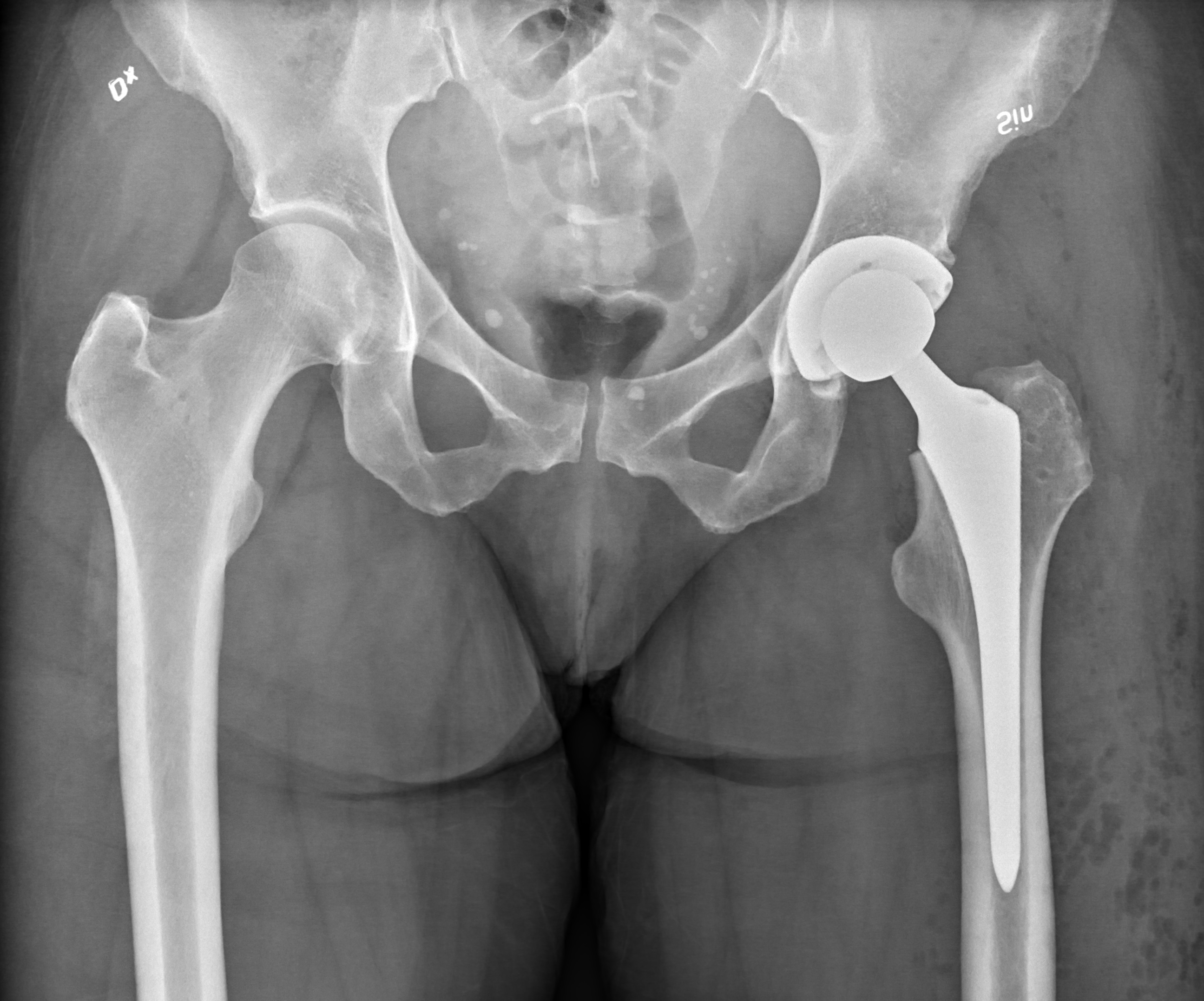
Here the prosthetic hip joint dark left can be seen with the peg implanted in the femur centre-left and replacement socket in the hip upper left.

Hip replacement x ray. A standard hip X-ray examination generally includes an anteroposterior PA image and a lateral image. Recovering from a hip replacement Staff on the ward will look after you when you come back from the operating theatre and you will usually be able to have something to drink within an hour or so after you get back to the ward. For example pelvic x-rays used for planning hip replacements vary in magnification sometimes from 115 to-130.
Then the hip muscles and tendons that were temporarily moved or cut to get into the hip joint are put back or repaired. This is an autoimmune disorder where the bodys immune system attacks its own tissue. This degree of variation occurs partly because of differences in techniques but mainly due to patients body habitus.
Product X-ray Images. Coloured X-ray of a mans pelvis front view showing a prosthetic hip implant red lower left. Follow-up X-rays are advised every two to three years after that for as long as the patient has the implant.
This X-ray shows the bones in your hip and gives a good idea of the health of the joint. It is used for the assessment of unilateral hip pathology most commonly to diagnose a hip fracture or dislocation. The implant is attached by a peg into the femur bone green with surgical cement.
At left is an X-ray of a patients hip with rheumatoid arthritis. X-ray images taken before and after a hip replacement operation. X-ray for hip arthritis explained To aid diagnosis of arthritis in the hip joint a doctor may arrange for you to have a hip X-ray.
Here the prosthetic hip joint dark left can be seen with the peg implanted in the femur centre-left and replacement socket in the hip upper left. Radiography is the primary imaging method for the evaluation of hip arthroplasties and imaging of a hip arthroplasty and its complications primarily relies on the information that is. Information gained from the initial radiograph includes assessment of the quality of implantation and hence the likelihood of long term success.
The lateral direction may be opted for in axiolateral images or a frog leg lateral image. Coloured X-ray of a section through the pelvic region of a 66-year-old male patient after total hip replacement surgery. The authors of this instructional course for orthopedic surgeons recommend routine monitoring starting five years after total hip arthroplasty THA or replacement.
Ideally the AP image shows both hip joints which strictly speaking makes it a pelvis X-ray to allow comparison with the other hip. SOLUTION SYSTEM Revision Hip System. Right This x-ray of an arthritic hip shows severe loss of joint space.
In 2006 over 55000 primary total hip replacements were implanted in the UK. S-ROM Modular Hip System. This is a basic article for medical students and other non-radiologists A hip x-ray also known as a hip series or hip radiograph is a pelvis x-ray with an additional lateral view of the specified hip.
The socket centre left of the ball-and- socket joint which is part of the pelvis bone has also been replaced. Left In this x-ray of a normal hip the space between the ball and socket indicates healthy cartilage. X-ray of a hip replacement in a 50 year old woman - hip replacement xray stock pictures royalty-free photos images Joey McLeisterStar Tribune MinneapolisMnFriAug.
A hip X-ray can often show the presence of arthritis in the joint. DePuy Synthes Revision Solutions. The right joint is affected by rheumatoid arthritis.
Coloured X-ray of a section through the pelvic region of a 66-year-old male patient after total hip replacement surgery. X-ray scan image of hip joints with orthopedic hip joint replacement implant head and screws in Hip joint replacement xray. You will have an X-ray before being discharged to make sure that your hip replacement looks normal.
X-ray of hip replacement to ensure it. Showing ball and socket joints titanium implant in medical orthpodedics scan. The head of the thigh bone femur forms a ball which fits into a socket in the pelvic bone.
A crucial aspect of follow-up for these patients is the assessment of the postoperative radiograph. Deciding to Have Hip Replacement. X-ray Right hip showing hip replacement or hip prosthesis made from titanium X-ray of the prosthesis of the left hip joint.

Main Complications Of Hip Arthroplasty Pictorial Essay
Hip Replacement Wikipedia
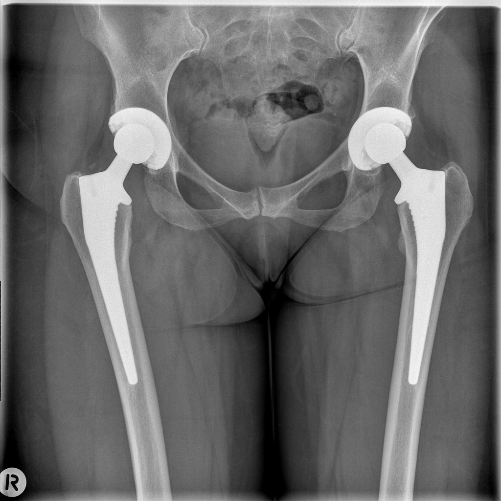
Normal Pelvis And Both Hips With Bilateral Total Hip Replacements Radiology Case Radiopaedia Org
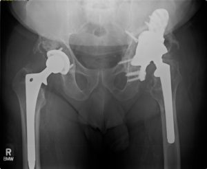
Revision Total Hip Replacement Richmond Va Dr Wind Orthova

X Ray Scan Image Of Hip Joints With Orthopedic Hip Joint Replacement Or Total Hip Prosthesis On Right Side Implant Head And Screws In Human Skeleton In Blue Gray Tones Movement Orthopedics